Blog

January 7, 2023

Tripping, Stumbling or Knocking feet? Proprioception explained.

There are several key areas to address when training for optimal performance. These include strength, endurance, power, balance and proprioception. I hear you, you’re probably thinking, ‘What on earth is that last word?’ Also known as kinesthesia, many people don’t know what proprioception is, how to train it, or how it helps with performance. We are here to explain! 

Proprioception is essentially the bodies perception of where and how our joints and limbs move in space. This sensation arises from the input of tiny sensory receptors that are located within your muscles, tendons and skin tissues. These signals travel to the brain, telling the body what movements need to occur, with what forces, and at what particular point in time. Additionally, it combines with other senses (Vestibular, nervous and vision) to locate external objects relative to the body and contributes to body image (Haan et al., 2015). For example, proprioception allows you to walk without thinking about each single step, it also allows you to touch your elbow with your eyes closed. If you had poor proprioception, you may trip or stumble frequently, or knock into doors as you walk past. Proprioceptive acuity has been found to be significantly correlated with high level sporting acheivements in elite athletes (Haan et al., 2015). Well-developed proprioception largely contributes to efficient strength, balance, co-ordination and correct movement patterns, ultimately improving you (or your horse) as an athlete. 

When there is injury to any tissue, for example a muscle tear, not only do the muscle fibers become disrupted, but so do these very small sensory receptors within it. This impairs the normal feedback the brain receives from this area, subsequently effecting the muscles' ability to accurately produce force, the timing of its activation and its functional role. Additionally, lack of proprioception may occur more globally within one's body, for example, after having several months of rest and de training. 

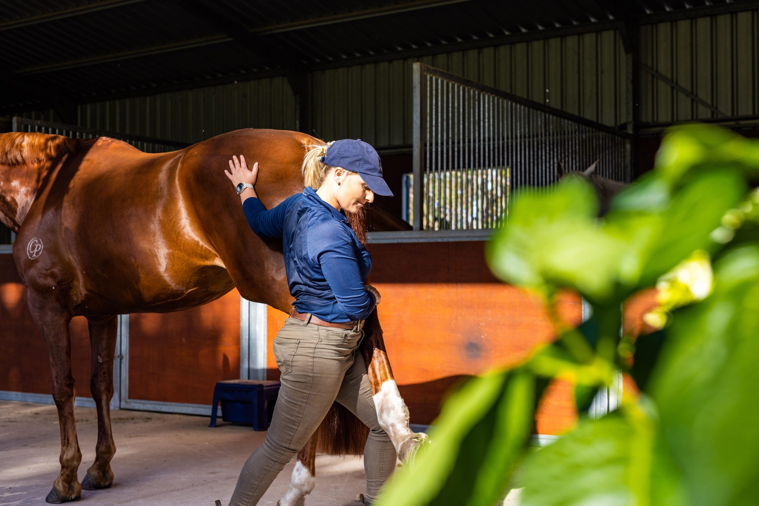
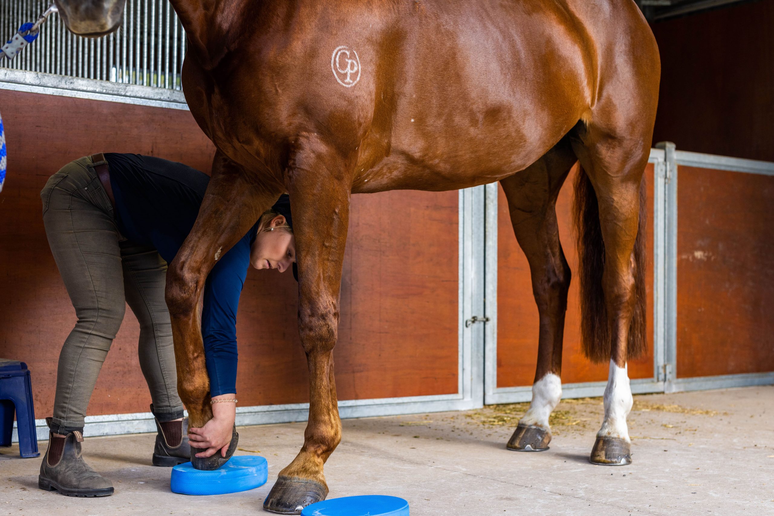
Physiotherapists are experts in physiological rehabilitation, and can provide a specific and individualized exercises to target proprioception in an area that may have been injured or weak, or out of practice. 

If your horse is often knocking their feet on each other, intermittently stumbling or tripping, or simply have or have had an injury, it may be worth seeking some physiotherapy input to improve proprioception and therefore performance. 

For the rider, if you find you slouch to one side, or often have uneven rein contact your physiotherapy may be able to help you with some proprioceptive posture coaching. Asymmetrical positioning and guidance can interfere with the aids from the rider when performing exercises, resulting in miscommunication, thereby negatively affecting performance and welfare of the horse. A recent study by Heidbuchel et al (2022) indicated that experienced riders who have better bodily proprioception experience decreased rein contact variability, increased postural control and a more balanced seat than novice riders on an equine simulator. 

References 

Han, J. et al. (2015) “Level of competitive success achieved by elite athletes and multi-joint proprioceptive ability,” Journal of Science and Medicine in Sport, 18(1), pp. 77–81. 

Heidbuchel, A. et al. (2022) “Assessment of kinematics on an equestrian simulator and physical fitness according to different skill levels of dressage riders,” SSRN Electronic Journal

January 7, 2023

Tendon load management… A fine balance between too much and too little.

Tendons are firm, usually cylindrical or flat structures that connect muscles to bones. They are made up of collagen, which has strong but elastic properties allowing shock absorption and transfer of forces. Under load, tendons undergo a stretch shortening cycle, where they lengthen, building and storing energy which is released as physical energy when the tendon shortens again. Think of a slinky, which uses repeatable stretch shortening cycles to create movement. Or a kangaroo, hopping around. Tendons act in similar ways in horses, providing economy for gait and force generation of the limbs, which are controlled by muscles closer to the center of their body. 

Gait analysis and electromyographic studies have shown that a horse's use of tendons in this way actually reduces the energy required by the horse by up to 36% during gait (O’Brien, Marr and Thorpe, 2020; Verkade, Back and Birch, 2019). 

These studies have also shown us activities and phases of gait that experience the highest loads, such as landing from a jump, and on mid stance during gallop (O’Brien, Niel and Marr, 2020). Another factor that increases tendon load is duty factor (the amount of time in contact with the ground), which is increased in high level dressage movements such as passage (Murray et al., 2006). Other factors that increase tendon loading include increase in gait speed, and more compliant, softer surfaces such as sand (Crevior-Denoix et al., 2013). 

Tendon injury occurs when there is repeated and extensive stretching of tendons during training. This overwhelms the capacity of tendon cells to repair before the subsequent loading cycle, leading to disruption to the collagen organization, which reduces the tensile strength of the tendon and makes it more susceptible to future overload (Bohm, Mersmann and Arampatzis, 2015). Disrepair and micro tearing within the tendon then generates a pain response, causing dysfunction and poor performance. 

Human and equine tendons are comprised of identical structural and cellular make up, allowing the transference of decades of tendon management knowledge from humans, to the equine animal. In order to improve function, the first and foremost thing to address is pain. To do this, physiotherapists around the world use slow, heavy isometric (Static) exercise to provide pain relief and pain free stimulation to tendon structures, and this concept can be applied exactly the same in horses (Bohm, Mersmann and Arampatzis, 2015). 

Once pain is controlled and the tendon has been de-loaded adequately, it is time to begin a progressive and graded strengthening and load management program. Qualified physiotherapists are able to evaluate the amount of load that is enough to generate cellular adaptation while avoiding overload and microdamage. Doing this stimulates collagen growth and organization in the tendon, laying down structure once again to allow its function of shock absorption and storage of energy (Heinemeier and Kjaer, 2011. 

If your horse has a tendon injury, your physiotherapy will work together with you over a period of months (Tendons have longer remodeling time frames than other tissues) to ensure the correct and appropriate load for the stage of recovery (Bohm, Mersmann and Arampatzis, 2015). References 

Bohm, S., Mersmann, F., & Arampatzis, A. (2015). “Human tendon adaptation in response to mechanical loading: a systematic review and meta-analysis of exercise intervention studies on healthy adults”. Sports medicine - open, 1(1), pp. 7. 

Brumitt, J., & Cuddeford, T. (2015). “Current Concepts of Muscle and Tendon Adapation to Strength and Conditioning”. International journal of sports physical therapy, 10(6), pp. 748– 759.

Crevier-Denoix, N. et al. (2013) “Comparison of superficial digital flexor tendon loading on asphalt and sand in horses at the walk and trot”. The Veterinary Journal, 198. 

Firth, E. (2006) “The response of bone, articular cartilage and tendon to exercise in the horse”. Journal of Anatomy, 208(4), pp.513-526. 

Heinemeier, K. and Kjaer, M. (2011) “In vivo investigation of tendon responses to mechanical loading', Journal Of Musculoskeletal & Neuronal Interactions, 11(2), pp. 115-123.

Lichtwark, G., Watson, J., Mavrommatis, S. and Wilson, A. (2009) “Intensity of activation and timing of deactivation modulate elastic energy storage and release in a pennate muscle and account for gait-specific initiation of limb protraction in the horse.” Journal of Experimental Biology, 212(15) pp.2454-2463. 

O’Brien, C, Marr, N, Thorpe, C. (2021) “Microdamage in the equine superficial digital flexor tendon.” Equine Vet Journal, pp. 53: 417– 430. 

R.C. Murray, S.J. Dyson, C. Tranquille, V. Adams (2006) “Association of type of sport and performance level with anatomical site of orthopaedic injury diagnosis - Equine Exercise Physiology.” Equine Veterinary Journal, pp. 411-416. 

Verkade, M., Back, W. and Birch, H. (2019) “Equine digital tendons show breed-specific differences in their mechanical properties that may relate to athletic ability and predisposition to Injury.” Equine Veterinary Journal, 52(2), pp.320-325. 

Wilson, A., Watson, J. and Lichtwark, G. (2003) “A catapult action for rapid limb protraction.” Nature, 421(6918), pp.35-36.

January 7, 2023

Is my horse naughty, or trying to tell me something?

I am constantly amazed at the willingness of most horses, and the amount of discomfort they generally put up with before they will let us know. Whilst there is always the possibility of a horse with a naughty pre-disposition, more often than not when a horse displays significant behavior, they are actually trying to tell us something. It is easy to get trapped in the routine of saying ‘he always does this’ or ‘just ride him through it’. Without the ability for verbal communication, It is absolutely essential for horse owners to be aware of how facial expression, behaviors and movement restrictions may indicate discomfort or pain. By being in tune with your horse, recognizing and respecting certain behaviors that appear unusual, you may be able to address small issues before they become larger problems. There is large benefit in pro-actively assessing and monitoring our horses and looking out for the signs, rather than waiting for them to become in enough pain to be obviously lame. Knowing how to read your horse and recognize signs of pain is particularly important in preventing significant injury, and moreover remains our responsibility as owners to instill welfare throughout management and performance activities involved with husbandry and horsemanship. Additionally, being aware of signs of pain or discomfort are also a good way to understand how your horse responds to increase in exercise load or a new activity.

Is your horse particularly naughty to pick out his hind hooves, or shoe on a particular hind limb? Perhaps he has significant stability or pain issues relating to a sacroiliac dysfunction. Is your horse girthy and snakey to tack up? Perhaps he has a really sore back from a poorly fitted saddle. Or maybe he flinching or ‘hates his ears being touched’… he is potentially experiencing a raging headache from a neck related issue. These signs of behavior certainly don’t mean that your horse has ‘this issue’, but it should flag to you that something could be causing discomfort, and may need to be screened by your qualified health practitioner. Horses are best to interact with and perform best when they are feeling good. Therefore, it is in your best interest to ensure you horse remains comfortable and free of pain.

Facial expressions have long been used in human health to effectively assess a range of painful conditions, from mild to severe pain. Similarly, (Mullard et al 2017) developed an equine ethogram which has been found and effective and practical method in detecting equine pain as well as determining the efficacy of the methods we use to ameliorate pain in horses. If your horse is beginning to display symptoms similar to these, you may need to consult your equine health practitioner.

This ethogram covers 24 behaviors listed below, of these, 20 have a strong correlation to lameness (Mullard et al., 2017). If horses exhibit approximately 8 or more behaviors, it is highly indicative that this horse is experiencing musculoskeletal pain (Mullard et al., 2017). One study which investigated correlations between ethogram scores and 5-star eventing performance found a significant relationship between low ethogram scores and final placings (Dyson and Ellis, 2020). There were moderate correlations between dressage penalty scores and ethogram scores and horses that failed to complete the cross-country phase had higher scores compared with those that completed (Dyson and Ellis, 2020).

  • Repeated changes of head position not in rhythm with trot
  • Head tilted
  • Head in front of vertical > 10s
  • Head behind vertical >10s
  • Tossing head
  • Ears rotated back behind vertical or flat >5s
  • Eyelids closed 2-5 seconds or frequent blinking
  • Sclera (white of eye) exposed repeatedly
  • Intense stare or glazed expression >5s
  • Mouth opening/separation of teeth repeatedly for 10 seconds
  • Tongue exposed protruding or hanging out
  • Tail clamped to middle or carried to the side
  • Tail swishing large and repeated movements during transitions
  • Rushed gait, irregular rhythm
  • Hindlimbs do not follow tracks of forelimbs
  • Wrong canter lead or disuniting
  • Spontaneous changes of gait i.e breaking from canter
  • Stumbles or trips more than once, toe dragging
  • Sudden change of direction against rider cues (Spooking)
  • Reluctance to move forwards
  • Rearing
  • Bucking or kicking backwards
    (Mullard et al., 2017)
    References
    Dyson, S. and Ellis, A.D. (2020) “Application of a ridden horse pain ethogram to horses competing at
    5‐Star three‐day‐events: Comparison with performance,” Equine Veterinary Education, 34(6), pp.
    306–315.
    Dyson, S. and Pollard, D. (2021) “Application of the ridden horse pain ethogram to elite dressage
    horses competing in World Cup Grand Prix competitions,” Animals, 11(5), p. 1187.
    Mullard, J. et al. (2017) “Development of an ethogram to describe facial expressions in ridden horses
    (fereq),” Journal of Veterinary Behavior, 18, pp. 7–12.
    Dalla Costa, E., Stucke, D., Dai, F., Minero, M., Leach, M.C., Lebelt, D. (2016) “Using the Horse Grimace
    Scale (HGS) to Assess Pain Associated with Acute Laminitis in Horses (Equus caballus).” Animals.
    6(8):47. https://doi.org/10.3390/ani6080047
    Dyson, S. and Van Dijk, J. (2018) “Application of a ridden horse ethogram to video recordings of 21
    horses before and after diagnostic analgesia: Reduction in behaviour scores,” Equine Veterinary
    Education, 32(S10), pp. 104–111.
December 21, 2022

Foot balance, Your Farrier and Physiotherapy.

3- Dimensional static and dynamic foot balance is essential for neutral and evenly dispersed loading through from the hoof, distal limb and into the center of the horse. Farriers (both barefoot trimming and shoeing) undergo significant training in their profession and are the experts when it comes to ensuring not only hoof care but the foundations for a musculoskeletally sound animal. Farriers are able to modulate grip, slide, leverage and load by taking into account hoof wall, coronet and digital angles, natural conformation, weight, and hoof tissue quality among other factors. Using physics, they can trim and/or shoe your horse to optimize distribution of ground reaction forces, balanced propulsion, appropriate digital cushion and hoof perfusion. 

There are important musculoskeletal structures (deep and superficial digital flexor tendons + Navicular bone) that insert or interact with the distal pedal bone, located deep within the hoof and pastern. Therefore, any deviation from the ideal point of balance, or center of rotation has the potential to put these structures and others under increased or imbalanced loading patterns. Additionally negative palmar angles in the distal limb places increased eccentric loading on the SDFT and DDFT, subjecting them to potential injury and overload. 

Not only do distal limb structures become affected by hoof shape and balance, but a flow on effect of compensation occurs higher up simultaneously. For example, Dr Kilmartin (2014) outlined an example of the implications along the front limb myofascial lines that occur in response to hoof imbalance. He stated that even a small amount of imbalance can cause a change in muscle development and tension in the upper body, “In cases of medio-lateral imbalance in one of the forelimbs where the medial wall of the hoof is more vertical, and the lateral wall is flaring out. Looking at the sole of the hoof the medial wall is higher than the lateral wall. In these cases, the Transverse, Ascending, and Descending Pectoral muscles are working along with the Subscapularis and Brachiocephalic to keep the fore limb under the body. These horses again consistently show pain or reactivity over the cartilage of the scapula.” This statement demonstrates the potential postural compensations a horse may make as a consequence of poor hoof balance. 

Additionally, Dr Ridgway (2016) describes an animal’s response to imbalanced propulsive forces due to high-low hooves, “The horse has to twist his head and neck to keep the eyes level. Horses, therefore, often exhibit muscle pain, stiffness, and spasm at the base of the neck.” This is why it is important to work with both your qualified farrier and physiotherapist to ensure a balanced, and healthy system from the hoof up. 

If you are seeking further information, www.theequinedocumentalist.com is an excellent resource for those wanting to learn more about the relationship between hoof balance and the wider musculoskeletal system.

Design by Insil
Follow us:
arrow-right